Figure 1: Endoparsite slides and description
courtesy of IDEXX RADIL
Protozoa (flagellates)
 |
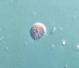
Slide 1: shows photo of the cyst and trophozoite form of Spironucleus muris. Note the teardrop shape of the trophozoite and its flagella (there are six anterior and two posterior). |
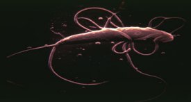 |
Slide 2: shows photo of electron micrograph of Spironucleus muris. Again note its shape and flagella |
 |
Slide 3: shows photo of differential interference contrast (DIC) photo of a Giardia muris trophozoite. Note the two anterior nuclei (“eyespots”) which give the organism a “monkey face” appearance. |
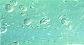 |
Slide 4: is a DIC photo of the trophozoite of Tritrichomonas muris. Note its lemon shape (Tritrichomonas sp. also have a characteristic undulating membrane and three flagella; these features are not evident on this slide but should be apparent on wet mounts). NOTE: Tritrichomonas sp. do not form true cysts. |
Pinworms
 |
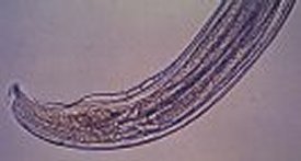
Slide 1: is a photo of a female Syphacia muris, the rat pinworm. Note the location of the vulva (slightly caudal to that of Syphacia obvelata). The vulva os Syphacia muris is located in the anterior fourth of the body. |
 |
Slide 2: is a photo of a tape test of a rat infected with Syphacia muris. Note the shorter slightly banana-shaped ova as compared to ova of Syphacia obvelata. |
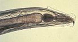 |
Slide 3: is a photo of Aspiculuris tetraptera, another rodent pinworm. Note the characteristic oval esophageal bulb and prominent cervical alae. |
 |
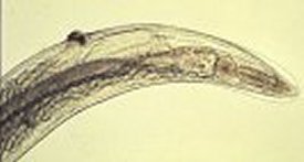
Slide 4: is a photo of the tail of a male Aspiculuris tetraptera.
Note the blunted point. |
Nematodes
 |
Slide 5: is a photo of a direct smear of urine from a rat infected with Trichosomoides crassicauda, the rat bladder worm. |
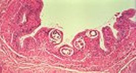 |
Slide 6: is a photo of a histologic section of a bladder from a rat infected with Trichosomoides crassicauda. Note the sections of worms within the bladder mucosa. |
Cestodes
 |
Slide 7: is a differential interference contrast (DIC) photo of the head of an adult Hymenolepis nana. Note the four prominent suckers and “armed” rostellum. |
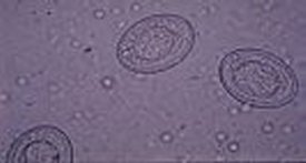 |
Slide 8: is a photo of an egg of Hymenolepis nana. Note the three pairs of hooks. |
Cestodes (intermediate host)
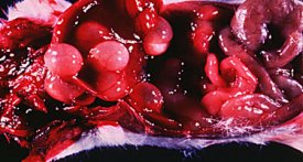 |
Slide 9: is a photo of a mouse infected with Cysticercus fasciolaris, the intermediate form of the cat tapeworm Taenia taeniaformis. Note multiple cystic structures attached to the liver. |
Slides and descriptions courtesy of:
DISEASES OF RESEARCH ANIMALS-DORA University of Missouri (Comparative Medicine Program and IDEXX-BioAnalytics)

